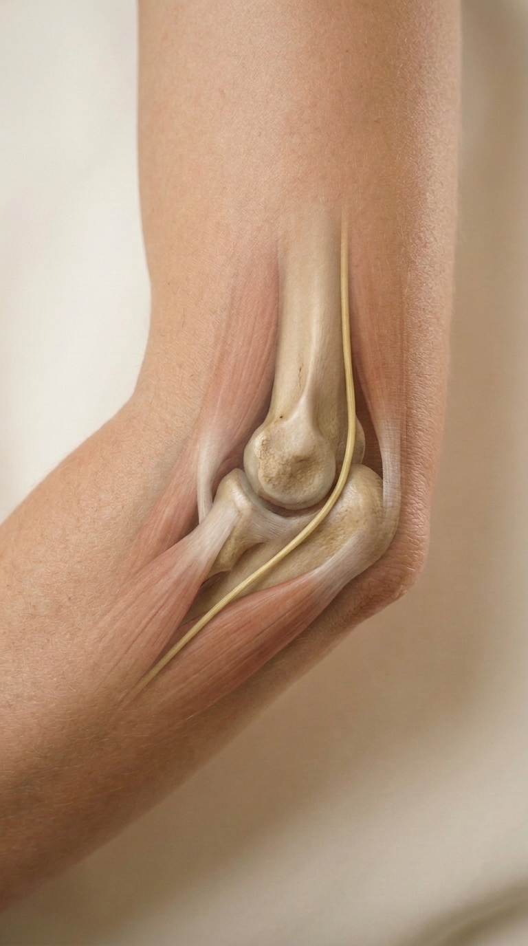
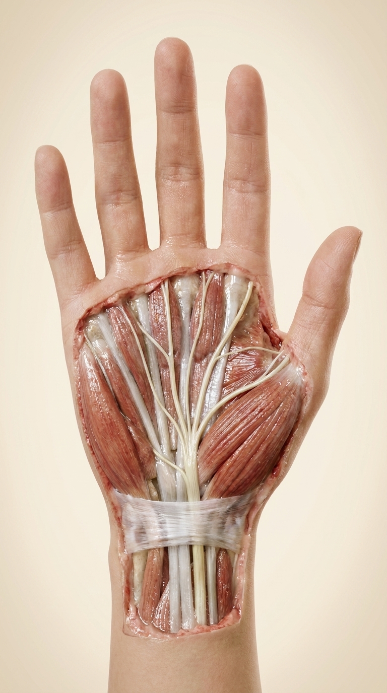
Matched end frame for the posteromedial RIGHT elbow photograph in reference 2. Reference 1 is for lighting only. Exactly match reference 2 elbow flexion, silhouette, magnification, joint position and warm cream background. Show a small educational virtual dissection confined to the posteromedial elbow. Softly fade overlying skin and fascia in a localized oval, continuous gradient into surrounding intact skin. True-to-life tissue colors and longitudinal texture. Accurately identify the medial epicondyle of the distal humerus and the olecranon of the proximal ulna as TWO distinct landmarks. One thin pale straw-yellow fasciculated ulnar nerve follows the medial distal upper arm, curving posterior to the medial epicondyle in the groove BETWEEN the medial epicondyle and the olecranon, then descends distally beneath the aponeurotic arch between the humeral and ulnar heads of flexor carpi ulnaris, disappearing into muscle. The cord lies alongside bone, not through it and not across the anterior crease or on the tip of the olecranon. Keep muscle attachments intact. No stray branches, labels, arrows, glowing lines, instruments, wound or lifted skin. Photorealistic clinical atlas, anatomically proportionate. Show a near-identical camera framing.